Parallel Projections Between Visual Cortical Areas
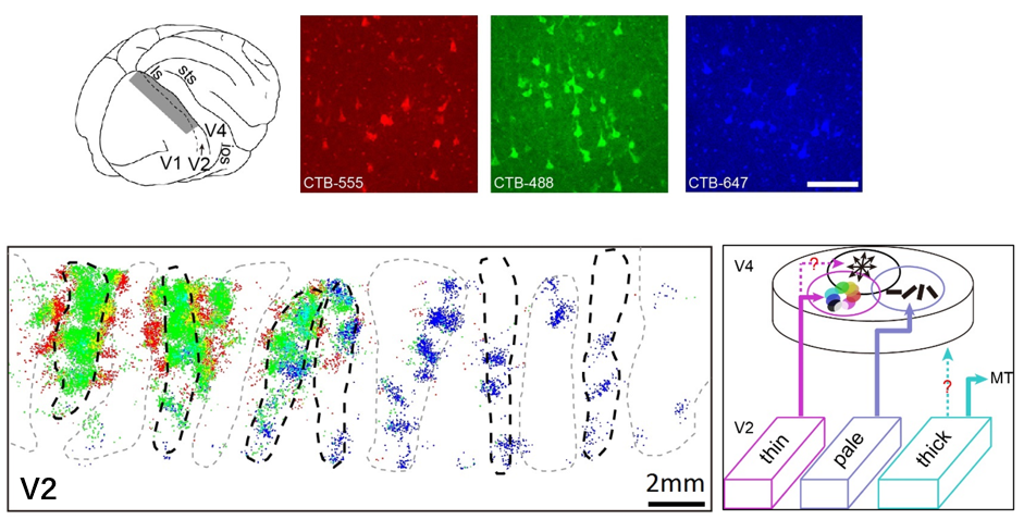
In key primate visual areas V2 and V4, functional modules exist for processing color, orientation, and motion information. However, it remained unclear whether parallel, functionally specific axonal projections connect these modules. The laboratory of Haidong Lu employed intrinsic signal optical imaging to functionally map orientation-, color-, and motion-selective domains in macaque V4, then injected differently colored retrograde tracers (CTB conjugates) into each domain.
They discovered that retrogradely labeled V2 neurons—marked by distinct fluorescent tracers—were segregated into different cytochrome oxidase (CO) stripes with remarkable spatial separation. Specifically: Tracer injections in V4 color domains labeled neurons in V2 thin stripes. Injections in V4 orientation domains labeled neurons in V2 pale stripes
By analyzing the correlation between functional specificity at V4 injection sites and the stripe distribution of labeled cells in V2, they established a projection blueprint: V2-to-V4 connections maintain parallel pathways for color and form information. Notably, no analogous connectivity pattern was observed between motion-processing modules of the two areas.
This study provides the first evidence of segregated parallel projections between functionally matched domains in V2 and V4, demonstrating that visual information is processed through both integrated and relatively independent parallel circuits. These findings fundamentally advance our understanding of information flow in visual cortical hierarchies (Fang et al., 2022).


 Address:No.138, medical college road
  Postcode:200032
  Tel/Fax:021-54237056
 Emial:itbr@fudan.edu.cn