Intrinsic Signal Optical Imaging
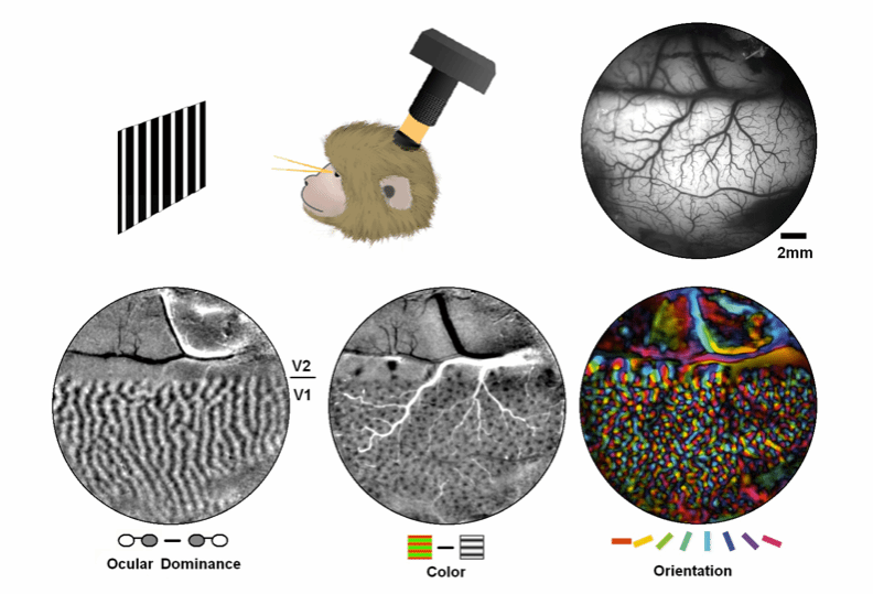
The figure demonstrates cortical vasculature and three representative functional maps recorded from the visual cortex of a macaque using intrinsic signal optical imaging (Lu et al., 2010). Intrinsic signals refer to hemodynamic activity that does not rely on exogenous agents (e.g., fluorescent dyes). Similar to near-infrared imaging, this method measures changes in reflected light intensity to assess neural activity in the cortex and compares responses to different stimuli to derive functional architecture (functional maps) within the cortical tissue.
Intrinsic signal optical imaging offers high spatial resolution (~0.1 mm), making it a crucial technique for studying functional organization at submillimeter scales. The intrinsic signal typically peaks 1–2 seconds after neural activation, but under certain conditions, it can achieve a temporal precision of around 0.1 seconds (Lu et al., 2017). This method has also been applied in intraoperative functional imaging studies during human brain surgery.


 Address:No.138, medical college road
  Postcode:200032
  Tel/Fax:021-54237056
 Emial:itbr@fudan.edu.cn